AI Fetal Imaging
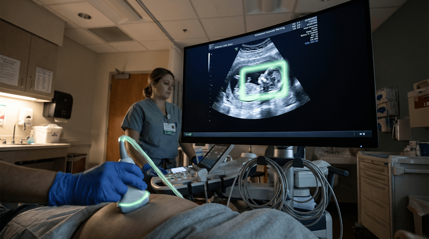
AI Fetal Imaging represents a significant advancement in prenatal diagnostic technology, employing deep learning algorithms to analyze ultrasound video streams during routine obstetric examinations. The technology works by training convolutional neural networks on vast datasets of annotated fetal ultrasound images, enabling the system to recognize anatomical structures, measure biometric parameters, and identify potential developmental anomalies. These algorithms process ultrasound data in real-time, automatically detecting features such as the nuchal translucency, cardiac chambers, neural tube development, and limb formation. The system can also track fetal growth markers including biparietal diameter, femur length, and abdominal circumference, comparing measurements against standardized growth curves to flag potential concerns. By leveraging pattern recognition capabilities that surpass human visual processing in certain contexts, these AI systems can identify subtle variations in tissue density, organ structure, and blood flow patterns that might otherwise go unnoticed during manual examination.
The implementation of AI-assisted fetal imaging addresses several critical challenges in prenatal care delivery. Traditional ultrasound interpretation depends heavily on operator skill and experience, leading to significant variability in diagnostic accuracy, particularly in resource-limited settings or regions with shortages of specialized sonographers. Research suggests that AI systems can help standardize diagnostic quality across different healthcare facilities, reducing the dependency on individual expertise while supporting less experienced practitioners in making more accurate assessments. This technology also addresses the time constraints faced by busy obstetric practices, as automated measurement and preliminary screening can accelerate examination workflows, allowing clinicians to focus on complex cases requiring human judgment. Furthermore, these systems can help reduce the anxiety associated with waiting for specialist review by providing immediate preliminary assessments, though final diagnostic decisions remain under physician oversight.
Early deployments of AI fetal imaging systems have appeared in both academic medical centers and commercial prenatal care settings, with several platforms receiving regulatory clearances in various jurisdictions. The technology is being integrated into existing ultrasound equipment through software updates or as standalone analysis platforms that process captured images. Current applications focus primarily on second-trimester anatomy scans, where the technology assists in the systematic evaluation of fetal organs and structures required by standard screening protocols. Industry analysts note growing interest from healthcare systems seeking to expand access to quality prenatal care, particularly in underserved areas where telemedicine-enabled ultrasound combined with AI interpretation could bridge geographic gaps in specialist availability. As the technology matures, future developments may extend beyond anomaly detection to include predictive analytics for pregnancy complications, integration with genetic screening data, and three-dimensional reconstruction capabilities that provide more comprehensive fetal assessments. This trajectory aligns with broader trends toward precision medicine in maternal-fetal care, where data-driven insights enable earlier intervention and more personalized management strategies throughout pregnancy.